Percutaneous Tracheostomies in Intensive Care: Insights and Best Practices
by Dr. Elliott Bertram-Ralph. MBChB BSc FRCA FFICM. ICM/Anaesthesia Consultant at Wythenshawe Hospital, Manchester, UK.

Elective tracheostomies in the intensive care unit are performed in approximately 10% of patients requiring mechanical ventilation for longer than seven days.1 Before the recent COVID-19 pandemic, this represented approximately 12,000 to 15,000 insertions per year in the UK.2 Two thirds of new tracheostomies are performed in intensive care units in patients with medical rationale for admission.3
The indications for a tracheostomy in intensive care include:
- Prolonged respiratory failure to enable weaning from mechanical ventilation (usually greater than 7–10 days).
- Upper airway obstruction.
- Access for pulmonary toilet and removal of bronchial secretions.
- Inability to protect airway and protection from aspiration.
The optimal timing of tracheostomy insert is a controversial topic, with limited data available to guide clinicians. It is important to weigh the benefits of continued trans-laryngeal ventilation versus the tracheostomy procedure. Endotracheal tubes are unpleasant to tolerate, require heavier sedation, and may cause trauma and damage to the vocal cords.
Existing medical literature supports three clinical benefits of tracheostomies.
- Improved patient comfort
- Earlier discharge from the ICU
- Shorter length of stay in the ICU11
Tracheostomies are also thought to support significant psychological progress for patients by aiding communication and comfort, reducing the risk of vocal cord damage, and allowing sedation to be weaned.
Surgical vs Percutaneous Tracheostomy Techniques
There are two main types of tracheostomy: percutaneous, which will be the focus of this summary, and surgical. The surgical technique involves careful dissection and identification of tissue planes, and creation of a stoma. It allows for better control of bleeding and is associated with fewer intra-operative complications. The percutaneous method involves the Seldinger technique to guide blunt dilators over a guidewire to create a stoma for the tracheostomy to be inserted through. It has been shown to be associated with fewer post-procedural complications, reduced tissue trauma’ and consequently a reduced chance of bleeding.
Percutaneous tracheostomies were first introduced in 1955 by Sheldon et al as an alternative to the surgical route. The progressive dilational technique was introduced by a surgeon from New York called Pat Ciaglia in 1985, where he used a modified percutaneous nephrostomy set to perform the tracheotomy. This percutaneous technique has been associated with reduced post-procedural complications such as bleeding and infection. Furthermore, no transfer is required of an unwell patient to the operating room, with generally improved cosmetic effects. Additionally, PDT is found to be cheaper and more efficient than surgical techniques. With evolution over time, the percutaneous tracheostomy technique has reduced the need for surgical tracheostomies.4
The absolute contraindications for percutaneous tracheostomy include i.a. patient refusal, localised infection, and uncontrolled coagulopathy. Relative contraindications include challenging ventilation and high oxygen requirements. Additionally, difficult airways or anatomy (for example, obese patients, enlarged thyroids, an unstable c-spine, or previous radiotherapy) should be considered.
The Percutaneous Tracheostomy Procedure Explained
I am a senior registrar in intensive care and anaesthetics, and I have gained most of my experience inserting tracheostomies over the past 12 months during my last year of training. Having more familiarity with the procedure has given me more confidence managing them day to day. Initially, the procedure seemed a daunting task. However, I found learning from expert colleagues who have kindly supervised me and utilising resources such as the National Tracheostomy Safety Project to be helpful. The method I describe below is based on my clinical experience.
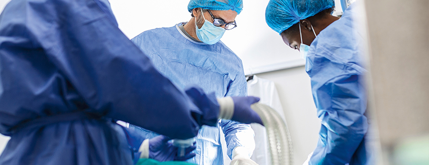
The percutaneous tracheostomy is the highest risk elective procedure that happens in the intensive care unit. 90% of ICU tracheostomies are performed percutaneously at the bedside.10 The Ciaglia method means that a minimum of three people are needed at the bedside in a carefully planned procedure. One person is required to manage the sedation and monitor and respond to observations. The second person is required to manage the airway and utilise a bronchoscope to allow the procedure to be performed under direct vision. The final person is responsible for inserting the tracheostomy. With several people conducting the procedure, human factors are important to consider; appropriate introductions and roles should be established before the procedure, and checklists and algorithms should be used to ensure safe patient care.
Adequate sizing is important for weaning, to allow air to bypass the tracheostomy for production of voice while reducing the pressure required to inspire and expire sufficient tidal volumes through the tracheostomy. I have found in my experience that generally a size eight tracheostomy is used for larger individuals and a size seven for smaller adults.
I would check blood tests prior to the procedure, ensuring coagulation is appropriate. The airway of the patient should also be assessed to ensure that the endotracheal tube can easily be replaced orally if required, with the difficult airway trolley on standby through the procedure. The patient should be appropriately monitored, e.g. with end tidal capnography throughout. Starvation status should also be fitting, and the NG can be aspirated before the procedure. Consent is mandatory, and if necessary, relatives should be informed, and patients appropriately supported to facilitate consent.
The patient should be easy to ventilate with minimal oxygen requirements. From experience, good ventilatory compliance and reduced dependence on positive end expiratory pressure allows a procedure to be completed more safely. Patients suitable for a tracheostomy ideally should be on less than 50% oxygen with a PEEP under 10cmH20 and haemodynamically stable with low vasopressor requirements.
I have always scanned the neck prior to the procedure by ultrasound to ensure that the tracheostomy can be safely inserted, for example, to review the depth of the trachea from the skin and to assess if there are any large midline blood vessels that could significantly increase bleeding risk*. Once scanned, local anaesthetic with adrenaline can be administered subcutaneously to ensure that the patient is comfortable and bleeding risk is further reduced. The position of the patient is also crucial; slight head up positioning can reduce venous pressure and bleeding, the neck can be extended by placing a pillow under the shoulders of the patient.
The person managing the airway should consider placing the patient onto 100% oxygen, altering the ventilator settings appropriately for a paralysed patient, and positioning them safely for the neck extension required for easy access to the trachea. Sedation should be increased so the patient is appropriately anaesthetised for the procedure and neuromuscular blockade. Then, under direct vision using a laryngoscope (a videolaryngoscope often makes this easier), the endotracheal tube with a deflated cuff should be withdrawn so the cuff is visible at the cords. At this point the cuff should be reinflated under direct vision and ventilation should be re-established. Once this has been completed, a bronchoscope is used to view the trachea from the proximal end. In my experience, having someone hold the tube in place reduces the risk of tube dislodgement.
The person Inserting the tracheostomy should be fully scrubbed and have prepared all required equipment. The neck should be cleaned with appropriate solution and a large drape used to cover the patient. Often two drapes are needed, one for the patient’s neck and face (ensuring easy airway access) and the second drape over the body and bed, providing a large field for aseptic technique. The curved single tapered large dilator can be placed in water at this stage, which allows activation of the hydrophilic coating to aid its passage through the tissue.
Once the neck has been adequately cleaned and local anaesthetic with adrenaline administered, a small safety scalpel can be utilised to provide a small and shallow incision to the skin. After this, you can use a gloved finger or some surgical dilators to open the tract slightly more. You should be able to feel the trachea and have a good impression of the midline. Often the light at the bronchoscope tip can be visible in the trachea at this point which provides a clear site to aim for.
There are many approaches to carrying out dilation and subsequent insertion, dependent on equipment. Here is an example of a step by step guide using a Tracoe Experc.
Potential complications and considerations
There are risks associated with PDT including malposition, worsening ventilation or oxygenation, and damage to local structures such as nerves (e.g vagus, recurrent laryngeal), the thyroid gland, blood vessels, and the trachea. Bleeding is reported in 5% of tracheostomies, for example puncturing an aberrant anterior jugular vein causes significant bleeding. The risk of death from the procedure itself is low, but ultimately the one-year mortality rate is around 37%.7
In my experience, a midline insertion is important to prevent uneven pressure of the tracheostomy on the mucosa leading to ulceration. There is also potential for loss of a patent airway during the procedure. An unsecured airway can lead to decruitment in the more PEEP dependent patients resulting in desaturation. Inadvertent extubation, bronchospasm and aspiration pneumonitis can all occur.
Posterior tracheal wall injury occurs more commonly in elderly patients who have thinner tracheal walls. This has the potential to cause subcutaneous emphysema or a pneumothorax., Oesophageal injury is rare, but can occur when excessive force is used. Although the minimally traumatic insertion system aims to reduce the risk of traumatic injury.
Inadvertent tracheal ring fracture is frequently missed at the time of occurrence. This can lead to tracheomalacia in the long term, where the trachea becomes less rigid and can collapse under negative pressure ventilation, causing airway obstruction.9
After the Insertion: Post-Operative Care and Follow-Up
A chest x-ray can be ordered post-operatively to assess for complications, although there is evidence a chest x-ray is only necessary after difficult procedures, to avoid exposure to radiation and save on costs.5 Other important aspects for patient safety include reviewing sedation and ventilation after the procedure, and production of a sign above the bed with details of the tracheostomy including when it was inserted and the type of tracheostomy. The National Tracheostomy Safety Project produce a useful bedhead sign to use.6 The cuff pressure should also be checked every 8-12 hours with a target of between 20-25mmHg with no leak.
After the procedure, the stoma will take around 7-10 days to mature and be considered established. The first change of the tracheostomy should be done with the utmost care, with a plan for difficulty during re-insertion. Most patients who have a percutaneous tracheostomy on the intensive care unit will only need it for a short period of time. The median time a tracheostomy remains in situ is 28 days, with hospital stays in these patients lasting around 50 days (with wide variations).9 When a patient is ventilator free and has had their cuff deflated for 24 hours, decannulation can be considered. This should involve careful MDT assessment to ensure this can be done safely.
If the patient is discharged with a tracheostomy from the ICU, it’s important they are placed in an area of the hospital that has the skills to manage it and a tracheostomy passport should be produced detailing key aspects, for example the reason of insertion, schedule for changes, and plan of future care. All patients who have had tracheostomies should be followed up, with details given to the general practitioner. ICU follow-up clinics also have an important role in reviewing and assessment of potential problems during recovery. Failure of stoma healing, granulomas, tracheal stenosis, and tracheomalacia can occur and any symptoms should be investigated appropriately.
Conclusion: The Value and Risks of Percutaneous Dilational Tracheostomies in the ICU
Percutaneous dilational tracheostomies have a significant role for patients in the ICU with potential benefits including weaning from mechanical ventilation and improved comfort. However, they are not without risk and should be performed with caution. I have found that the simple insertion kits provided, guidance from senior clinicians, and various teaching resources have helped me with improving my technique inserting and managing tracheostomies in the intensive care unit.

Dr. Elliott Bertram-Ralph MBChB BSc FRCA FFICM is a ICM/Anaesthesia consultant at Wythenshawe Hospital, Manchester, UK.
References
1Mehta C, Mehta Y. Percutaneous tracheostomy. Annals of cardiac anaesthesia. 2017 Jan 1;20(Suppl 1):S19-25.
2McGrath BA, Wilkinson K. The NCEPOD study: on the right trach? lessons for the anaesthetist. British journal of anaesthesia. 2015 Aug 1;115(2):155-8. [last updated 2014; cited 2023.07.21]. Available from: https://www.ncepod.org.uk/2014report1/downloads/OnTheRightTrach_FullReport.pdf
3Namin AW, Kinealy BP, Harding BC, Alnijoumi MM, Dooley LM. Tracheotomy outcomes in the medical intensive care unit. Missouri medicine. 2021 Mar;118(2):168.
4Byhahn C, Westphal K, Zwissler B. Percutaneous Tracheostomy: Past, Present, and Future Perspectives. InYearbook of Intensive Care and Emergency Medicine 2005 2005 (pp. 30-38). Springer New York.
5Tobler Jr WD, Mella JR, Ng J, Selvam A, Burke PA, Agarwal S. Chest X‐ray after Tracheostomy Is Not Necessary Unless Clinically Indicated. World journal of surgery. 2012 Feb;36(2):266-9.
6Paulich S, Cook TM, Hall H, Churchill H, Kelly FE. Two new algorithms for managing tracheostomy emergencies on the ICU. British Journal of Anaesthesia. 2020 Jul 1;125(1):e164-5. [last updated 2020; cited 2023.07.21]. Available from: https://www.tracheostomy.org.uk/NTSP-Algorithms-and-Bedheads
7Namin AW, Kinealy BP, Harding BC, Alnijoumi MM, Dooley LM. Tracheotomy outcomes in the medical intensive care unit. Missouri medicine. 2021 Mar;118(2):168.
8Yartsev, A. Complications of Percutaneous and Surgical Tracheostomy. Deranged Physiology; 2020. [last updated 2024.02.03; cited 2023.07.21]. Available from: Complications of percutaneous and surgical tracheostomy | Deranged Physiology
9McGrath BA, Wallace S, Lynch J, Bonvento B, Coe B, Owen A, Firn M, Brenner MJ, Edwards E, Finch TL, Cameron T. Improving tracheostomy care in the United Kingdom: results of a guided quality improvement programme in 20 diverse hospitals. British journal of anaesthesia. 2020 Jul 1;125(1):e119-29.
10Faculty of Intensive Care Medicine. Guidance for: Tracheostomy Care. ICS; 2020. [last updated 2023.08; cited 2023.07.21]. Available from: https://www.ficm.ac.uk/sites/ficm/files/documents/2021-11/2020-08%20Tracheostomy_care_guidance_Final.pdf
11Angus D, Finfer S, Gattioni L, Singer M. Oxford textbook of critical care. Oxford University Press; 2019 Dec 3:376.
12Zouk AN, Batra H. Managing complications of percutaneous tracheostomy and gastrostomy. Journal of Thoracic Disease. 2021 Aug;13(8):5314.