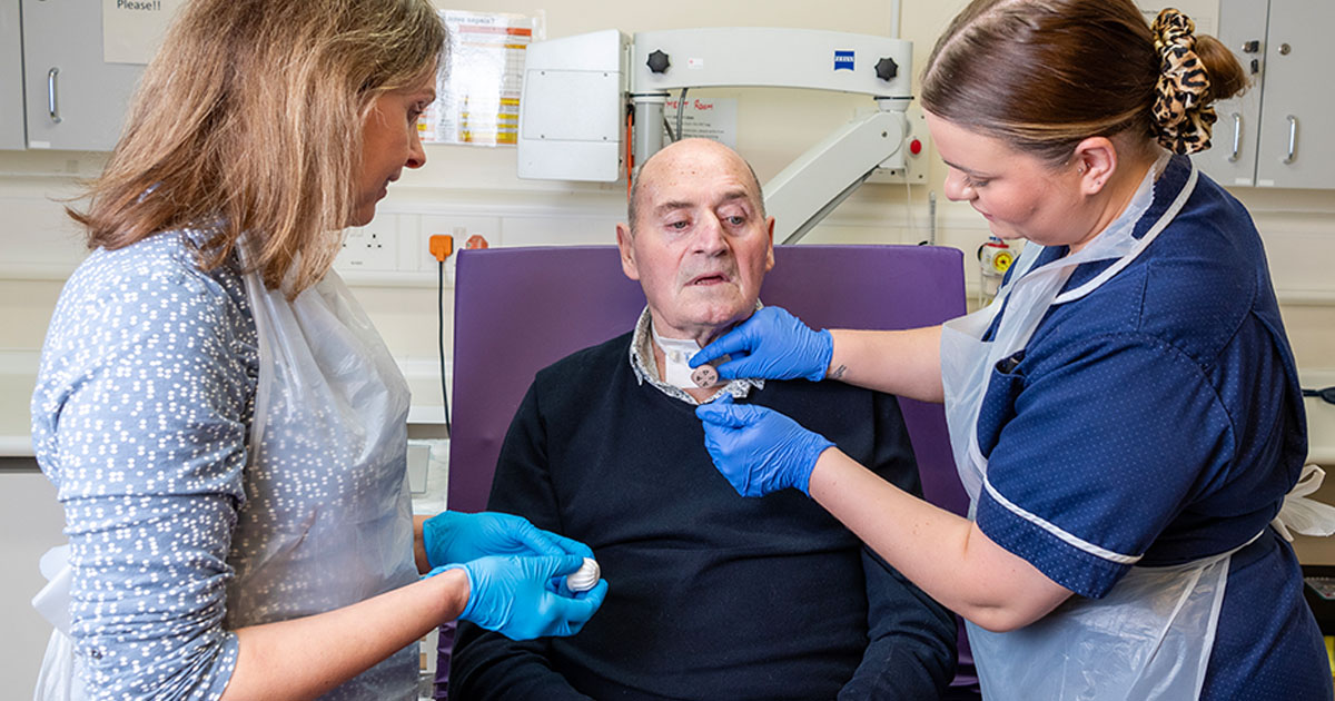
Diagnostic and Therapeutic Methods for the Decannulation of Tracheotomized Neurological Patients

Clinical Publications of Interest 2025-09
This edition highlights a comprehensive review of the decannulation framework of tracheotomized neurological patients, addressing the complex interplay of diagnostic and therapeutic approaches required for safe and effective weaning. Drawing on recent clinical research, the publication outlines key criteria, assessment tools, and interventions that support decision-making in intensive care and neurorehabilitation settings. The featured review by Dziewas et al. provides a structured protocol to guide clinicians through the challenges of dysphagia, airway management, and secretion control, with the goal of improving patient outcomes and reducing long-term complications.
Dziewas R, Warnecke T, Labeit B, Schulte V, Claus I, Muhle P, et al. Decannulation ahead: a comprehensive diagnostic and therapeutic framework for tracheotomized neurological patients. Neurol Res Pract. 2025;7(1):18.
Neurological patients in ICU often undergo tracheostomies, but decannulation is hindered by severe dysphagia
- Up to 47% of neurological patients in ICU, such as those with stroke, traumatic brain injuries and severe critical illness polyneuropathy, undergo tracheostomy.
- Patients often enter early rehabilitation while still cannulated due to the danger of weaning during the acute stage of treatment, however, decannulation is of great importance as permanent cannulation shows high mortality rates.
- Severe dysphagia and other factors such as impaired airway anatomy and reduced cough strength pose a serious challenge for decannulation.
Diagnostic procedures - factors to consider when determining readiness for decannulation
- Clinical examinations of swallowing and airway safety
- Cough strength and type/ amount of bronchial secretions
- Airway anatomy (stenosis)
- Decannulation criteria and scores - A2BC criteria
1. Clinical procedures such as the clinical swallow exam and Evans Blue dye test are used to assess swallowing, aspiration and penetration of food boluses and saliva
- The clinical swallow exam (CSE) has low reliability and sensitivity for determining swallowing safety.
- The Evans Blue dye Test (EBT) and modified Evans Blue dye Test (mEBT) have low negative, but high positive diagnostic value.
- Swallowing can be further assessed by the flexible endoscopic evaluation of swallowing (FEES), which can be performed at the bedside, allowing direct visualization of the swallow.
- The SESETD (Standardized Endoscopic Swallowing Evaluation for Tracheostomy Decannulation) protocol, established for the reliability of endoscopic exams, identifies four key diagnostic factors: extent and localization of salivary retentions, laryngeal sensitivity, subglottic and tracheal structures by transstomal endoscopy, and spontaneous swallowing frequency.
2. Quantitative measures of coughing and characterization of bronchial secretions serve as good indicators for safe decannulation
- The strength and effectiveness of secretion removal via coughing can be assessed with quantitative methods like peak cough flow (PCF) and maximum expiratory pressure (MEP).
- Qualitative clinical cough scores on a 6-point scale (the semiquantitative cough strength score) or by a binary test (white card test) are also employed.
- Semi-quantitative assessment of bronchial secretions, such as the modified semiquantitative airway score (m-sqAS), may involve measuring suctioning frequency and describing the characteristics of secretions, such as viscosity and color.
3. Airway stenosis is a common tracheostomy complication that is examined by endoscopic and manometric means
- Clinically relevant stenosis, both fixed and dependent, involving a lumen narrowing of at least 20%, is expected in 10-20% of patients.
- Capping, use of a speaking valve, and change of tracheostomy tube can be considered by measuring the intrathoracic pressure by tracheal tube manometry.
- The main treatment for stenosis includes optimizing the tracheal cannula design (ie. using smaller outer diameter) to minimize tissue damage and using corticosteroids for profound edema.
4. A2BC decannulation criteria link all the clinically relevant diagnostic domains for decannulation
- There are a multitude of decannulation criteria and scores used, with patient observation, clinical and endoscopic exams being common.
- Criteria for decannulation depend on the clinical setting and patient population: alternative criteria may need to be considered for neurological patients in ICU compared to patients in early neurorehabilitation in intermediate care.
- The A2BC criteria consider the best diagnostic procedures across relevant clinical domains and propose the most effective combination: airway safety according to SESETD protocol during FEES, airway anatomy by translaryngeal endoscopy, bronchial secretions by m-sqAS or suctioning frequency, and cough strength by spirometry. The specific thresholds, qualitative or quantitative, indicate if a patient is ready for decannulation.
Therapeutic interventions - how to bring patients closer to decannulation
- Physiological airflow in the upper airways
- Above-cuff vocalization
- Pharyngeal electrical stimulation
- Other - swallowing behavioral therapy, ice chip protocol, secretion management
1. Physiological airflow through the upper anatomy can be achieved by tracheal cannula weaning
- Gradual weaning requires progressively longer periods of cuff deflation accompanied by a closed tracheostomy tube or by use of speaking valve.
- Restoration of airflow through the upper anatomy improves pharyngeal and laryngeal sensation, resulting in better secretion management.
- Gradually increasing air resistance allows for strengthening of respiratory muscles.
2. Above-cuff vocalization (ACV) helps in laryngeal stimulation
- ACV is a feasible and safe method that allows tracheotomized patients to produce speech by directing controlled airflow over the vocal cords.
- The technique stimulates the larynx, which may improve swallowing function and facilitate the weaning process, though standardized protocols must be established.
3. Dysphagia can be successfully treated by pharyngeal electrical stimulation
- Pharyngeal electrical stimulation (PES) involves applying an electric current along a specialized feeding tube to the pharyngeal mucosa, with intensity being adapted to the patient.
- PES causes a reorganization of connections in the regions of the motor cortex associated with swallowing and increases sensory input to the swallowing circuitry via the periphery nerve endings.
4. Other diagnostic or therapeutic methods include swallowing behavioral therapy, ice chip protocol and secretion management
- Swallowing behavioral therapy targets swallowing with restorative and compensatory exercises as reduced swallowing activity can worsen dysphagia and secretion buildup.
- The ice chip protocol improves swallowing by producing a strong sensory response to ice chips and can be used as both a diagnostic and therapeutic tool.
- Secretions can be managed by anticholinergic drugs or botulinum toxin A injection to the salivary glands, as well as humidifying breathing air and endotracheal suctioning.
A newly developed framework for decannulation management of ventilator-weaned tracheotomized patients contains a fast and standard track pathway
- Assessment of contraindications, such as respiratory dependencies and acute infections, for evaluations such as cuff deflation must first be performed.
- If there are no contraindications, criteria according to A2BC are evaluated.
- The fast-track pathway involves assessment of A2BC criteria, which, if any criteria are not met, necessitates the use of targeted therapies and re-assessment at regular intervals. Passing of the A2BC criteria allows for decannulation.
- The standard-track pathway involves restoration of the physiological airflow in the upper airways by gradual increase of cuff deflation times and subsequent occlusion training, accompanied by continuous contraindication monitoring. Patient stability during 24-38 hours with an unblocked and closed cannula and final confirmation of airway patency allows for decannulation.
Clinical Publications of Interest
Our quarterly update highlights studies from peer-reviewed journals on head & neck cancer, laryngectomy, and tracheostomy.
Want to learn more?
- Read an interview with Rainer Dziewas, on decannulation, the framework developed and his thoughts on ACV [Tracheostomy Decannulation in Neurological Patients: Protocols, Challenges, and Clinical Insights]
- Learn more about Above Cuff Vocalization (ACV) [Above Cuff Vocalization (ACV): Improving Communication for Tracheostomized ICU Patients - Atos Medical]
- Join our On-demand webinar ‘ACV: An exploration of clinical practice’ [ACV: An exploration of clinical practice - Atos Medical]
- Read an interview with Anja Fischer, Speech-Language Pathologist, where she shares her clinical expertise on the use of ACV in tracheostomized patients [What is Above Cuff Vocalization in Tracheostomy Patients? - Atos Medical]
Other relevant publications of interest
- Bath PM, Woodhouse LJ, Suntrup-Krueger S, Likar R, Koestenberger M, Warusevitane A, Herzog J, Schuttler M, Ragab S, Everton L, Ledl C, Walther E, Saltuari L, Pucks-Faes E, Bocksrucker C, Vosko M, de Broux J, Haase CG, Raginis-Zborowska A, Mistry S, Hamdy S, Dziewas R; for PHADER Investigators. Pharyngeal electrical stimulation for neurogenic dysphagia following stroke, traumatic brain injury or other causes: Main results from the PHADER cohort study. EClinicalMedicine. 2020 Nov 10;28:100608.
- Calderone A, Filoni S, De Luca R, Corallo F, Calapai R, Mirabile A, Caminiti F, Conti-Nibali V, Quartarone A, Calabrò RS, Rifici C. Predictive Factors of Successful Decannulation in Tracheostomy Patients: A Scoping Review. J Clin Med. 2025 May 28;14(11):3798.
- Otto-Yáñez M, Monge G, Muñoz T, Segovia E, Villalobos P, Vera-Uribe R, Resqueti V, Fregonezi G, Torres-Castro R. Predicting Decannulation Success in Patients With Neurological Conditions: Development and Validation of the NEURODECANN Clinical Score. Nurs Health Sci. 2025 Sep;27(3):e70200.
- Muhle P, Suntrup-Krueger S, Burkardt K, Lapa S, Ogawa M, Claus I, Labeit B, Ahring S, Oelenberg S, Warnecke T, Dziewas R. Standardized Endoscopic Swallowing Evaluation for Tracheostomy Decannulation in Critically Ill Neurologic Patients - a prospective evaluation. Neurol Res Pract. 2021 May 10;3(1):26.
- Likar R, Aroyo I, Bangert K, Degen B, Dziewas R, Galvan O, Grundschober MT, Köstenberger M, Muhle P, Schefold JC, Zuercher P. Management of swallowing disorders in ICU patients - A multinational expert opinion. J Crit Care. 2024 Feb;79:154447.
- Hernández Martínez G, Rodriguez ML, Vaquero MC, Ortiz R, Masclans JR, Roca O, Colinas L, de Pablo R, Espinosa MD, Garcia-de-Acilu M, Climent C, Cuena-Boy R. High-Flow Oxygen with Capping or Suctioning for Tracheostomy Decannulation. N Engl J Med. 2020 Sep 10;383(11):1009-1017.
Atos Learning Institute Global Newsletter
Stay up to date with the latest articles, news, and insights in laryngectomy and tracheostomy care with the Atos Learning Institute Global Newsletter – published quarterly.
Sign up for the newsletter and stay connected! [Atos Learning Institute Global Newsletter - Atos Medical]
Disclaimer:
The content of the journal articles is the opinion of the article authors and does not necessarily reflect the opinion of Atos Medical AB nor any of its subsidiaries. By providing this material it is not implied that the articles nor its authors are endorsing Atos Medical AB or Atos Medical AB products. Nothing in this material should be construed as Atos Medical AB providing medical or other advice, making any recommendations or claims, and is purely for informational purposes. It should not be relied on, in any way, to be used by clinicians as the basis for any decision or action, as to prescription or medical treatment. When making prescribing or treatment decisions, clinicians should always refer to the specific labeling information approved for the country or region of practice.
Clinical Publications of Interest summaries of journal articles are not exhaustive. For full content, please see the actual publication.

Atos Learning Institute offers a wide range of learning solutions, including local market classroom-based training and customized learning programs, virtual instructor-led training and blended learning. The institute has a team of experienced trainers and experts who design and deliver the training programs and partner with subject matter experts.